
Module 03
Swan journey
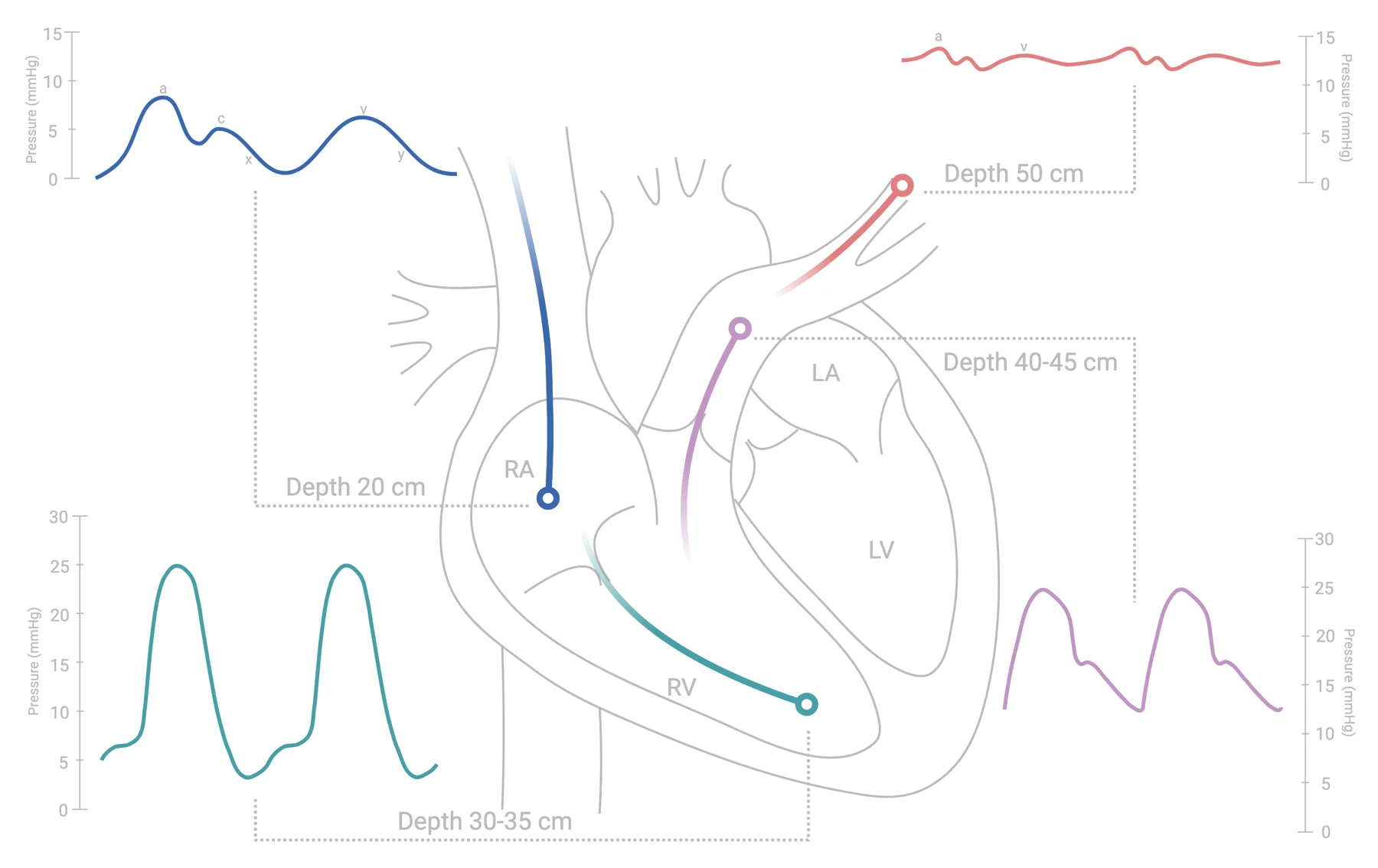
Float a pulmonary artery catheter from the right atrium to the wedge and watch the pressure trace change at each landmark. The depths are for a right internal jugular approach. Bootsma 2022 Kern 2022
Advance the catheter
Drag the depth, or let it float.
Patient physiology
The same journey looks different in disease.
RA mean
4mmHg
RV
25/4mmHg
PA
25/10mmHg
Normal: RV and PA systolic pressures match; PA diastolic ≈ wedge; RA mean ≈ RVEDP.
Superior vena cava / RA junction
14 cmAn atrial-type trace: small a, c and v waves; low pressure.
Normal
Mean 2–6 mmHg
At this position
Do not inflate the balloon until the tip is past the subclavian system; inflate once in the RA so the flow carries it.
Landmarks and what to do when you miss them
Right internal jugular approach
Which chamber is this?
The scale is hidden on purpose: read the shape, especially diastole.
Round 1/8
Best 0/8Diagnose by diastole
The fastest way to know which chamber you are in
- RA: small waves, low pressure, no systolic peak.
- RV: tall systolic peak, then diastole falls to near zero and slopes up to end-diastole (a-wave kick just before the next QRS).
- PA: similar systolic peak, but diastole stays up (≈ wedge level) and there is a dicrotic notch; pressure falls through diastole rather than rising.
- Wedge: back to an atrial pattern, damped and delayed, v ≥ a, mean below PA diastolic.
A thrombus in the lumen or a kink can turn a 70/35 PA trace into a convincing 45/20; when contours look damped, flush and re-check before believing the numbers.Nishimura 2012
Original figure
Damrongwatanasuk, Shan & Hu (manuscript in review)
Insertion essentials and safety
- Right IJ is preferred; ultrasound guidance cuts carotid puncture risk by about 70%. Catheter is 110 cm, marked every 10 cm, balloon 1.5 mL.
- Use fluoroscopy for severe RA enlargement, TR, RV dilation, a left IJ approach, or device leads.
- Zero to atmosphere; level at the phlebostatic axis (≈5 cm below the sternal angle). Above the axis under-reads; below over-reads.
- Read all pressures at end-expiration; average ≥3 beats; more in AF.
- Diagnostic catheterization complications in large series: death ≈0.2%, MI ≈0.05%, stroke ≈0.07%, serious ventricular arrhythmia ≈0.5%, major vascular ≈1%.